Diabetic patients, particularly older ones, may present with several diabetes-related ocular complications on a daily basis. As eyecare providers, we are uniquely qualified to not only manage the disease’s ocular complications but to also educate patients on how best to take care of themselves. Here, I discuss diabetes in the eye, the most up-to-date treatment.
Ocular Adnexa
Uncontrolled diabetic patients (those who do not check their blood sugar often enough and/or do not follow a strict regimen of diet and exercise) are at high risk for developing infections, such as preseptal and orbital cellulitis, vs. their controlled counterparts due to a weakened immune system.
Preseptal cellulitis presents as acute lid erythema and edema restricted to the soft tissues preceding the orbital septum. The bacterial infection is usually due to the local spread of dacryocystitis (nasolacrimal sac swelling), a neighboring sinusitis, from eyelid trauma, hordeolum or an external ocular infection. The most efficacious treatment is oral antibiotics.
Orbital cellulitis is characterized by a severely edematous painful upper eyelid, often accompanied by ophthalmoplegia, lid erythema, and fever. An afferent pupillary defect may be present depending on the amount of optic nerve involvement. This condition places patients at risk for meningitis and can lead to death. The management protocol for orbital cellulitis is an immediate referral to the nearest ER for stat CT, blood cultures and iv antibiotics.
Ocular Musculature
Uncontrolled diabetes, which is a microvascular disease, may cause poor circulation of the microvasculature that feeds the extra-ocular muscles. This can elicit ptosis secondary to levator weakening, as well as strabismus secondary to cranial nerve (CN) III, IV or VI paresis. Generally, CN VI tends to be more commonly affected at 50%, with CN III a close second at 43%. Cranial nerve IV is rarely affected at 7%, says a study in Ophthalmology.
When a diabetic patient presents with strabismus, consider the “whole” patient. If the patient is age 55 or older, the pathology is most likely microvascular. If the patient is younger than age 55, we need to rule out compressive lesions affecting the cranial nerves by having the patient perform a visual field test and referring him/her for a neurological evaluation.
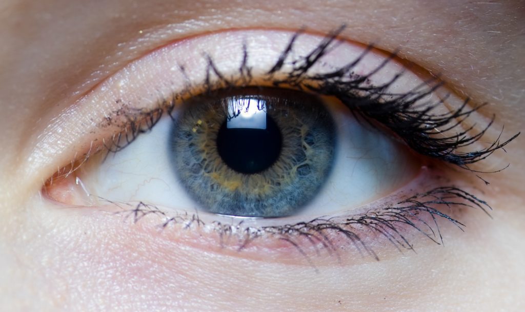
The Ocular Surface
Peripheral neuropathy, which is diabetes-induced nerve damage that causes loss of sensation, numbness and occasionally feet, leg or hand pain, can affect corneal sensitivity, interrupting the neurotransmission feedback loop. This, in turn, causes an increase in ocular surface inflammation and an elevated risk of dysfunctional tear syndrome. So, diabetic patients tend to have dry eye disease.
Treatment for these patients involves the aggressive use of preservative-free tears, as well as anti-inflammatory therapy (when inflammation is present) to alleviate any epithelial defects, which can place these patients at risk for keratitis. The tears can be used as much as necessary. Steroids are typically prescribed q.i.d., and cyclosporine ophthalmic emulsion 0.05% (Restasis, Allergan) or Lifitegrast 5% (Xiidra, Shire) is usually prescribed b.i.d.
Research reveals diabetic patients also exhibit a growing incidence of corneal epithelial pathologies, such as punctate epithelial erosions, recurrent corneal erosions, and corneal ulcers. The reason: Type 2 diabetics have an increase in sorbitol (a sugar alcohol the body slowly metabolizes), which can, in turn, damage the corneal epithelium. Specifically, increasing levels of glucose in the blood cause an increase in sorbitol, which induces a decrease in hemidesmosomes in the corneal epithelium. This reduction causes a weakened adhesion of the epithelium to the underlying stroma, leading to an increase in the breakdown of the epithelium.
For this reason, when selecting contact lenses for diabetic patients, prescribe strict daily wear, explain the importance of compliance on lens disposal, and educate on the significance of using efficacious cleaning and storage solutions. Also, direct these patients to immediately remove lenses if redness, discomfort, foreign body sensation or decreased vision occurs, as this may signal a serious infection.
Lens
In addition to the pathological changes in the cornea epithelium caused by an increase in sorbitol, this increase also induces an escalated rate of premature cataract formation. In addition, this sorbitol increase results in refractive error shifts secondary to the infusion of water into the lens matrix. Both hyperopic and myopic shifts are seen, but myopic shifts are more common. This is a frequent finding in newly diagnosed diabetic patients.
To best manage these patients, re-refract them four to six weeks after their first exam when their blood sugar is (hopefully) better controlled, to provide the most accurate prescription.
When co-managing cataract surgery patients, keep in mind that a long duration of postoperative anti-inflammatory therapy may be necessary to prevent complications, such as cystoid macular edema. Also, make sure the antibiotic drops are used q.i.d. for nine days post-op to prevent infections.
Vitreous and Retina
Non-proliferative diabetic retinopathy can be mild, moderate or severe. (See “Non-Proliferative Diabetic Retinopathy,” above.) Proliferative retinopathy is characterized by neovascularization in the retina and optic disc due to advanced capillary closure-caused ischemia. This occurs secondary to hypoxia in the posterior pole, causing an increased VEGF level. Neovascularization of the Iris (NVI), new vessels in the retina (NVE) or new vessels of the disc (NVD) are characteristic. These patients often present with decreased VA and/or floaters, secondary to vitreous hemorrhage.
PRP remains the mainstay treatment for proliferative retinopathy. The procedure reduces the need for oxygen in the posterior pole and subsequently decreases VEGF levels, thus preventing neovascularization. Steroid injections and implants have been shown efficacious in treating macular edema in these patients, and anti-VEGF medications injected directly into the vitreous cavity have proven successful as well.
Optic Nerve
The optic nerve is acutely at risk for damage in uncontrolled diabetes, secondary to its reliance on the microvasculature of the posterior ciliary artery. Uncontrolled diabetic patients are at a great risk for anterior ischemic optic neuropathy, retinal artery occlusion, retinal vein occlusion, worsening glaucoma, and diabetic papillitis. All these complications are worse with concomitant microvascular diseases, such as hypertension and hypercholesterolemia.
Our Role
In our yearly comprehensive examinations, we are able to detect and closely monitor ocular complications. This not only allows us to be the “gatekeepers” to our patients’ health, but it also enables us to be influential in their education. Motivating patients to monitor their blood sugar and maintain a healthy diet and exercise regimen may be one of our most important duties. One day, we may be able to influence a change from diabetes being the leading cause of blindness in the United States to a rare complication.