By Priyal Gadani, O.D., F.A.A.O.
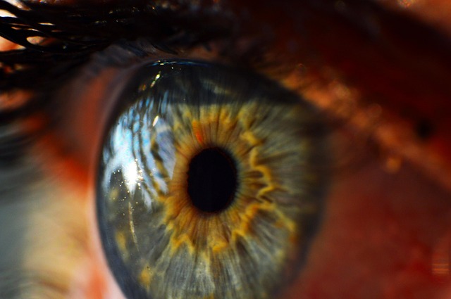
The cornea is the clear, dome-shaped outermost tissue of the eye, which is sometimes referred to as the window to the eye. The cornea focuses light through the lens onto the retina. A diseased or injured cornea can cause significantly decreased vision, pain, or discomfort. Oftentimes, these issues may be remedied by medication eye drops, contact lenses, or other more conservative procedures, but if the cornea still does not respond, a corneal transplant may be required.
A corneal transplant is a surgical procedure in which damaged or diseased cornea is replaced by donated corneal tissue (the graft). It may restore vision, reduce pain, and improve the appearance of the damaged or diseased tissue.
A number of conditions may be treated with a corneal transplant, including:
When the entire cornea is replaced, it is known as a penetrating keratoplasty and when only part of the cornea is replaced, it is known as lamellar keratoplasty. Keratoplasty is another term for surgery of the cornea. Penetrating keratoplasty (PK) involves a complete transplant of the cornea, and may be a better treatment option for patients with severe corneal disease. PK is also called a “full thickness” corneal transplant, meaning the entire cornea is removed and replaced with a donor cornea.
Corneal tissue used in corneal transplants come from recently deceased donors who have no history of known diseases or other factors that would affect the chance of success of the donated tissue or health of the recipient. Donors can be of any age.
Penetrating keratoplasty is an outpatient procedure, meaning that patients can go home the same day as their surgery. Before beginning the PK procedure, a local or general anesthesia is administered. The corneal surgeon uses a trephine, or circular cutting device, to cut the donor corneal tissue the exact size and shape needed. A second trephine is used to remove a circular portion of the patient’s cornea, after which the donor tissue is positioned and sewn into place. Antibiotic drops are applied and the eye is covered with a protective shield at the end of the procedure.
The patient will return the next day for a post-operative appointment with their ophthalmologist, and will use several medication eye drops to help prevent infection and inflammation. The vision will be blurry during the recovery process, and it may take several months for the eye to heal and vision to improve.
Initially, a protective shield is worn to protect the eye following the procedure, and in the following week and months, patients are asked to not rub the eye and avoid activities which may cause trauma to the eye such as playing sports.
Risks of PK are similar to those of other intraocular procedures including:
The body’s immune system may mistakenly attack the donor cornea in some cases—this is called rejection. Graft rejection, and detachment or displacement of the graft occurs in about 20% of cases. Graft failure can occur at any time after the cornea has been transplanted, even years or decades later.
Signs and symptoms of cornea rejection include:
If the first transplant is rejected, certain patients may need a second transplant. A repeat transplant carries a higher rate of rejection than the first.
The corneal transplant may be technically performed perfectly, and the transplant may be working as well as it can be, but other eye problems may limit the quality of a person’s vision post-operatively. The new cornea may have a significant amount of astigmatism and glasses or special contacts may be required to improve vision. Other eye diseases such as macular degeneration, diabetic retinopathy, or glaucoma may also limit the patient’s quality of vision and prevent the patient from seeing 20/20.
If you have a damaged cornea, corneal transplant may be a good option for improving and restoring clearer vision.
Make an appointment today at one of our eight convenient Atlanta-area locations.
Schedule an Appointment Online
Or call 678-381-2020